Notice: Trying to access array offset on value of type null in /home/u9176434/en.uretradarligi.com/wp-content/plugins/js_composer/include/autoload/vc-shortcode-autoloader.php on line 64
Notice: Trying to access array offset on value of type null in /home/u9176434/en.uretradarligi.com/wp-content/plugins/js_composer/include/autoload/vc-shortcode-autoloader.php on line 64
Notice: Trying to access array offset on value of type null in /home/u9176434/en.uretradarligi.com/wp-content/plugins/js_composer/include/autoload/vc-shortcode-autoloader.php on line 64
The Treatment of Urethral Stricture
The treatment of urethral stricture does not involve medications. It involves three surgical methods, which include dilation, endoscopic removal of the stricture (internal urethrotomy), and open surgery (urethroplasty). The selection of the method to be used depends on the cause, location, length, and severity of the stricture.
Endoscopic Surgery (Internal Urethrotomy)
Endoscopic surgery, also called internal urethrotomy, is performed when the stricture in the urinal canal is shorter than 2 cm. This surgery involves the use of a special device that carries light and a camera at its tip. This device is advanced through the urinal canal and the stricture is removed with a knife or laser light found at the tip of the device.
This surgery usually takes about one hour. Once the stricture is surgically relieved, a catheter is placed. This catheter remains for three days. After this catheter is removed, some patients are provided with special single-use catheters and asked to insert catheters themselves a few times every day. These catheters are removed after 10-15 minutes. The purpose here; is to prevent or delay the urinal canal, which has been relieved in closed surgery, from constricting again.
Following internal urethrotomy, the stricture in the urinal canal usually recurs and the patient may need to undergo another surgery. Closed surgery can be utilized for a maximum of two times. Thus, it should not be performed more than two times, because stricture length increases in parallel to the number of closed surgeries. The success rate of closed surgery varies between 35-70%. It is not recommended for pediatric cases of urethral stricture.
Open Surgery (Urethroplasty)
Open surgery is recommended in cases where closed surgery fails to deliver the expected benefits and where the stricture is longer than 2 cm. Open urethral surgery is known as urethroplasty.
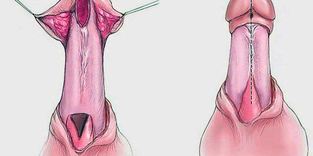
Various urethroplasty techniques exist. Urethroplasty may take up to 2-3 hours depending on the performed procedure. In this surgery, the stricture site is accessed through an incision made on the lower surface of the penis or immediately under the scrotum, depending on the location of the stricture in the urinary canal. If the stricture is shorter than 2 cm, this area is removed completely and the remaining healthy tissues are connected to form a new urinary canal. If the stricture is longer, this anastomosis method is not used. In such cases, a graft is made to the stricture site. The graft is obtained from penile skin or cheek mucosa. A catheter is left to remain for 2-3 weeks after surgery.
Some patients manifest a complete blockage of the urinary canal. The stricture in the urinary canal may not be treated with a single surgery in these patients. In individuals with complete blockage of the urinary canal without adequate tissue for a graft where the penis structure is unsuitable for the grafting procedure or anesthesia for long surgeries is risky, a temporary opening is created for the urinary canal immediately under the scrotum. A new surgery is performed six months after this procedure to move this urinary canal, which had been under the scrotum, to the tip of the penis. If the patient refuses to undergo a second surgery, the urinary canal may permanently remain under the scrotum. However, these individuals cannot urinate while standing up and have to sit down.
The rate of recovery of urinary canal strictures with open surgery is 90-95%.
Insertion of a Permanent Catheter into the Bladder Through the Abdomen
For patients who cannot be operated due to high anesthesia risk, an alternative option is to insert a permanent catheter into the bladder through a hole in the abdomen. This catheter is renewed at certain intervals and individuals can continue their normal lives. Conditions such as urinary tract infections, high fever, formation of bladder stones, and decrease in the volume of the bladder could be encountered due to the constant presence of a catheter.


